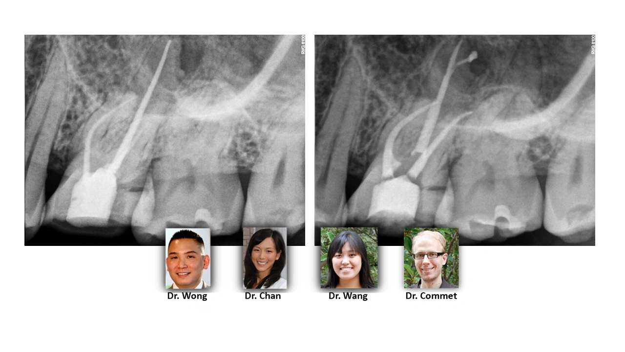
PRE-OPERATIVE ASSESSMENT

The pre-treatment radiograph shows several technical deficiencies in the existing obturation: (1) short DB fill and (2) missed MB2. Radiographically there is a lesion associated with a seemingly well filled palatal root. Careful inspection; however, demonstrates that this lesion is asymmetrically placed off to the distal. This guides treatment towards exploring for apical bifurcations. The post operative image shows that there was in fact an apical bifurcation filled with necrotic tissue. The unlocated MB2 and short DB fill were both corrected for, but it is likely that the patient’s symptoms and infection were attributable to this complex apical anatomy of the palatal root.