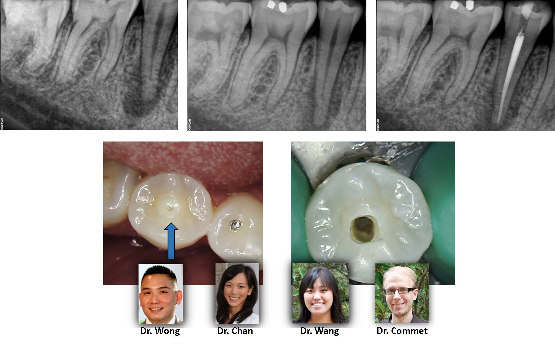
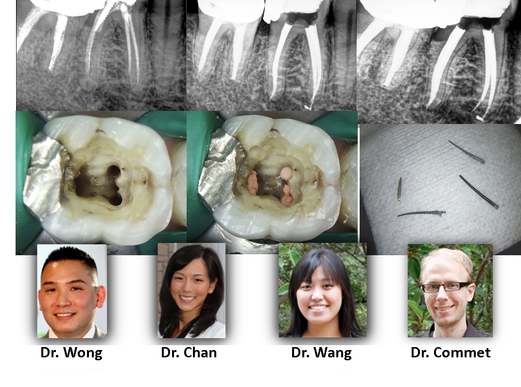
This tooth demonstrates the pitfalls of a dens evaginatus. As can be noted on the clinical photograph, the atypical central cusp quickly wears due to attrition resulting in pulpal exposure and corresponding necrosis. Careful inspection of the occlusal anatomy is important in these cases as the radiographic appearance is similar to that of a vertical root fracture but this tooth has a very favorable prognosis. […]
Fractured? Or just compromised?
This case was referred to our office for evaluation and expected extraction with eventual dental implant. Radiographically there was a large lesion associated with the mesial root, accompanying deep probing, and large swelling at the gingival margin. This case has several signs that would be consistent with non-restorable fracture. However, we used all available diagnostic tools, including CBCT, and could not definitively identify a fracture that would condemn the tooth to extraction. Therefore, we pursued retreatment with 6 weeks intracanal medicament. At the time of case completion, this patient was completely asymptomatic and the gingival tissue was healthy and firm. […]
ATYPICAL ANATOMY
This case represents a rare anatomic variant of a tooth anatomy much more commonly seen in the mandible: “c-shape”. Because the maxillary variant often has fused roots with DB/P canals that join very early in the pulp chamber with a separate MB canal that joins near the apex, some refer to this rare tooth as having a “semi-colon” anatomy. Take a look at the clinical photograph; can you see the semi-colon? […]
TOOTH RESORPTION:
This one is interesting. This patient winds up in our office for a 3rd opinion because the tooth had been condemned for extraction. After careful discussion with both patient and restorative dentist, we were able to identify that the resorption process was still above the level of the bone. Osseous crown lengthening was discussed as a potential requirement for an adequate restoration. We were able to completely degranulate the resorption tissue using the endodontic microscope, treat the area with acid, and repair with defect from within the endodontic access. Additionally, we placed a fiber reinforced post so that the restorative […]
Diagnosis:
Accurate treatment cannot happen without accurate diagnosis. This case was referred for evaluation and treatment of tooth #2. It was presumed that the large soft tissue swelling was coming from this tooth. As one can plainly see on the 2-dimensional radiograph, tooth #2 does have a lesion associated with its roots. Is this the cause of the large sized swelling in the soft tissue just behind it (clinical photo)? We used the CBCT to reveal complete occlusion of the right maxillary sinus secondary to sinus infection (purple *). The infection has traced the path of least resistance which happened to […]
Dental Anatomy 5
It is only Tuesday and here is a sampling of cases that have come through this week. We demonstrate bifurcations, middle mesial canals, radix entomolaris, significant curvatures. All of these are examples of anatomical considerations when rendering root canal therapy in order to maximize disinfection and long term success. Turkey Day is just around the corner!! […]
Rain or Snow!
Rain or snow doesn’t stop Renovo! Winter certainly had quite the kick off; we saw several inches of snow as well as some unusual cases. Despite the cold, our brave patients still made the trip to our office to visit! A rare root morphology is demonstrated by the three canal mandibular premolar (two more premolars on the contralateral side to come!) and an example of a four canal maxillary molar with internal coronal resorption that was repaired with composite after acid treatment. All in a good days work – only thing left is to hope for a reprieve from the […]
“Modern Endodontic Instruments”
With new heat treated NiTi files, we no longer need straight line access to achieve exceptional outcomes even in severely curved teeth. This is particularly important in cases like this, where the restorative dentist has already received the crown from the lab, and extreme care must be taken to provide as minimal of an access as possible in order to avoid taking new impressions and incurring additional lab expenses to remake the crown. […]
Cracked Tooth Syndrome…
Cracked Tooth Syndrome: Here is a case where our patient presented with significant pain with 2 teeth but neither the patient or referring Dr. could determine the root cause. Clinical objective testing MUST be done and proper diagnosis cannot be made by looking at an image alone. Significant findings included 1] deep isolate probing between the teeth but moreso when canted towards the posterior tooth (left one) 2] Periradicular bone loss around one of the roots causing a J-shaped appearance 3] Bone loss most extensive at the alveolar crest (green arrow), suggesting that the crack started at the level of […]
Retreatment…
Retreatment: Here is an interesting case of root canal retreatment from a failed initial attempt at initial endodontic treatment. This particular case was filled with a carrier-based filling material, which has no solvent. This, along with the presence of a metal post cemented in the tooth, makes it inherently more difficult to treat. If you look closely at the photo of the post-treatment access, there is a 3rd canal (not separate) within one of the roots. Another cool case! […]