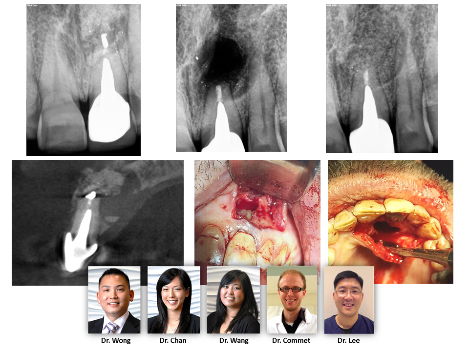
Here is a 79yo healthy female with non-remarkable medical history. She presented with a history of massive palatal swelling due to perforation of the palatal root from attempted retreatment by her dentist. As you can see, a part of the previous casted post remains in the palatal root. We have identified the perforation on 2D imaging and confirmed with CBCT (we area aware of an untreated MB2 canal; however, there is no evidence of periapical pathology and retreatment will not be attempted until we can confirm that we have rectifed the palatal pathology). We have decided to attempt repairing the […]
Category: Diagnosis
PRE-OPERATIVE ASSESSMENT
The pre-treatment radiograph shows several technical deficiencies in the existing obturation: (1) short DB fill and (2) missed MB2. Radiographically there is a lesion associated with a seemingly well filled palatal root. Careful inspection; however, demonstrates that this lesion is asymmetrically placed off to the distal. This guides treatment towards exploring for apical bifurcations. The post operative image shows that there was in fact an apical bifurcation filled with necrotic tissue. The unlocated MB2 and short DB fill were both corrected for, but it is likely that the patient’s symptoms and infection were attributable to this complex apical anatomy of […]
Fractured? Or just compromised?
This case was referred to our office for evaluation and expected extraction with eventual dental implant. Radiographically there was a large lesion associated with the mesial root, accompanying deep probing, and large swelling at the gingival margin. This case has several signs that would be consistent with non-restorable fracture. However, we used all available diagnostic tools, including CBCT, and could not definitively identify a fracture that would condemn the tooth to extraction. Therefore, we pursued retreatment with 6 weeks intracanal medicament. At the time of case completion, this patient was completely asymptomatic and the gingival tissue was healthy and firm. […]
TOOTH RESORPTION:
This one is interesting. This patient winds up in our office for a 3rd opinion because the tooth had been condemned for extraction. After careful discussion with both patient and restorative dentist, we were able to identify that the resorption process was still above the level of the bone. Osseous crown lengthening was discussed as a potential requirement for an adequate restoration. We were able to completely degranulate the resorption tissue using the endodontic microscope, treat the area with acid, and repair with defect from within the endodontic access. Additionally, we placed a fiber reinforced post so that the restorative […]
Diagnosis:
Accurate treatment cannot happen without accurate diagnosis. This case was referred for evaluation and treatment of tooth #2. It was presumed that the large soft tissue swelling was coming from this tooth. As one can plainly see on the 2-dimensional radiograph, tooth #2 does have a lesion associated with its roots. Is this the cause of the large sized swelling in the soft tissue just behind it (clinical photo)? We used the CBCT to reveal complete occlusion of the right maxillary sinus secondary to sinus infection (purple *). The infection has traced the path of least resistance which happened to […]
Cracked Tooth Syndrome…
Cracked Tooth Syndrome: Here is a case where our patient presented with significant pain with 2 teeth but neither the patient or referring Dr. could determine the root cause. Clinical objective testing MUST be done and proper diagnosis cannot be made by looking at an image alone. Significant findings included 1] deep isolate probing between the teeth but moreso when canted towards the posterior tooth (left one) 2] Periradicular bone loss around one of the roots causing a J-shaped appearance 3] Bone loss most extensive at the alveolar crest (green arrow), suggesting that the crack started at the level of […]