Cool Cases Archives | Page 2 of 5 | Renovo Endodontic Studio
An Unusual 'Post' Removal

AN UNUSUAL ‘POST’ REMOVAL

AN UNUSUAL ‘POST’ REMOVAL Apicoectomy can be a highly predictable treatment option for patients with failing endodontic therapy. Modern microscurgery involves not only root resection, but also retropreparation using ultrasonics and retrofil using bioceramic materials like MTA. Without these two key factors, treatment success drops from ~87% to nearly 60% and is the reason apicoectomy tends to have a “50/50” connotation with some oral surgeon offices. The following is a great case to demonstrate the possibilities in modern endo! Pre-operatively you can note that the ‘post’ (silver point) goes to the apex and CBCT showed a corresponding lesion. Placing bioceramics on […]

Read More… from AN UNUSUAL ‘POST’ REMOVAL

Broken Instruments...or Modified Root Filling?

BROKEN INSTRUMENTS…OR MODIFIED ROOT FILLING?

BROKEN INSTRUMENTS…OR MODIFIED ROOT FILLING? Here we present a relatively interesting case, #14, with a recurrent infection associated with the MB root. Other than pain associated with this tooth, medical history remains unremarkable. We were able to remove the “root filling” which appears to be endodontic instruments that may have also been used in initial instrumentation. In any case, we were able to remove the all three instruments and locate a previously untreated MB2 canal. This would prove to be significant as this it the only canal in the MB root that was able to be instrumented to full root […]

Read More… from BROKEN INSTRUMENTS…OR MODIFIED ROOT FILLING?

Endodontic Surgery...

ENDODONTIC SURGERY…

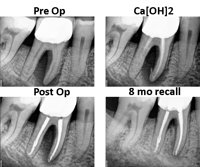
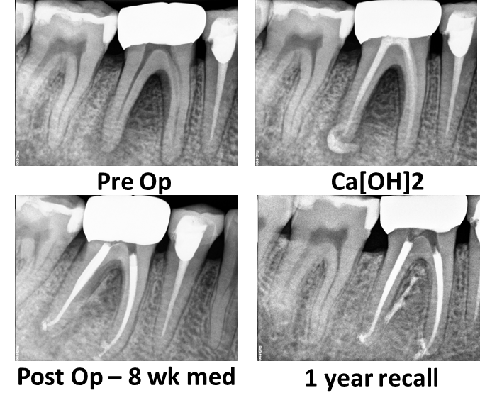
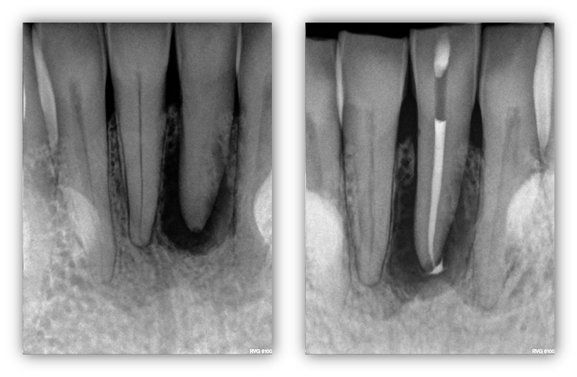
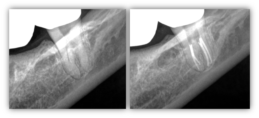
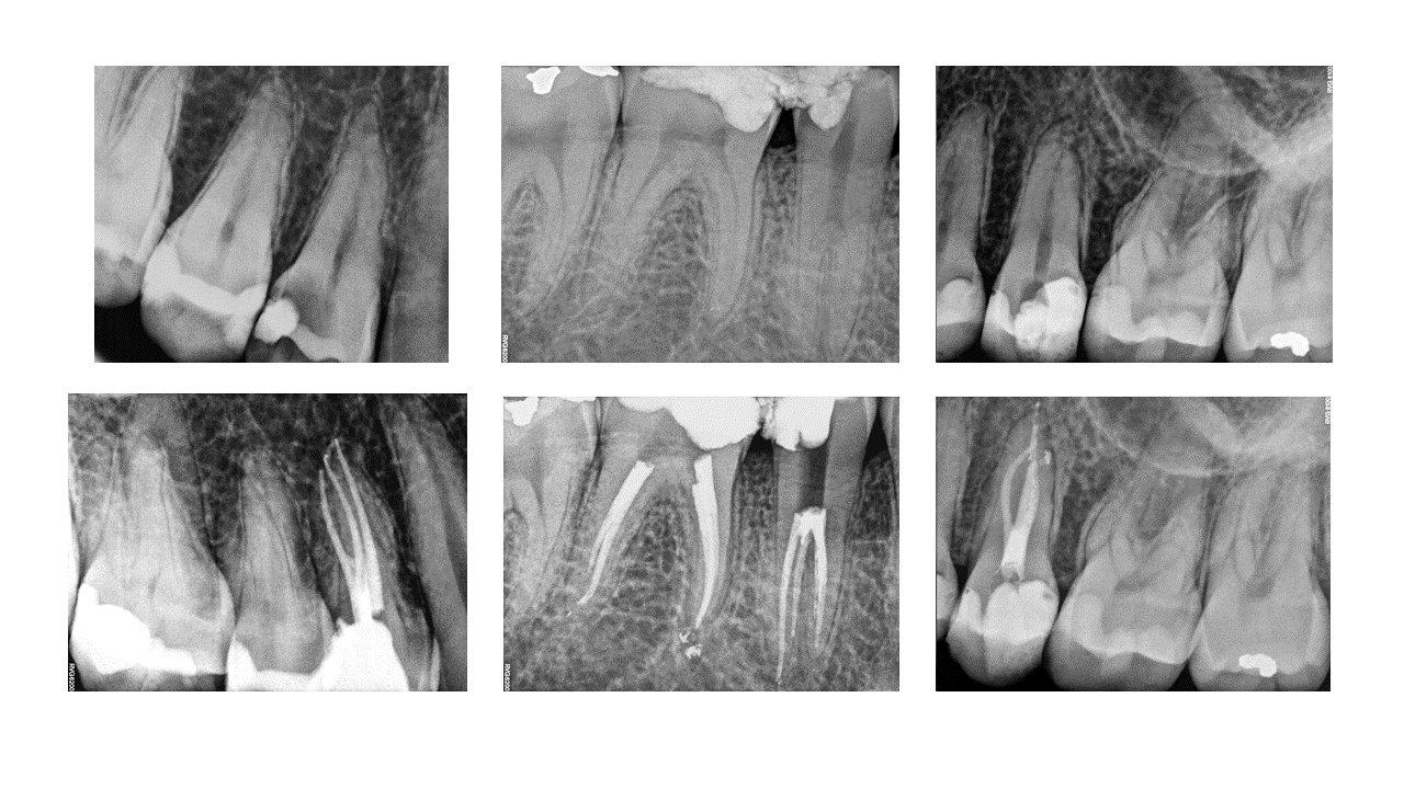
ENDODONTIC SURGERY: Here we present our patient, 33yo health female, who presented with a swelling in the mandibular central area. Root canal treatment when rendered when the patient was an adolescent. Years later, a periapical lesion developed with what appears to be a resorption defect that was not filled at the time of the original fill. We decided to so apicoectomy with conventional root end resection and retrofill, cortical allograft and resorbable membrane. As one can see, the entire buccal surface of the tooth was exposed due to the pathology. Upon post surgical follow-up and secondary recall, the surgical area […]

Read More… from ENDODONTIC SURGERY…

Endodontic Healing Potential

ENDODONTIC HEALING POTENTIAL 2

ENDODONTIC HEALING POTENTIAL: Here we present a case of the potential of healing disease with proper endodontic treatment. Pre-operatively the case displayed a large distal root lesion with significant furcation involvement, buccal swelling and probing that could essentially reach the root apex. Without any definitive proof of non-restorable tooth/root fracture, including pre-operative CBCT, we decided to initiate root canal treatment with the intent of saving the tooth. Upon the initial appointment the case was completely instrumented and medicated for 8 weeks (no sponge/cotton pellet used). At the completion appointment, we had already noted significant lesion healing with no clinical signs […]

Read More… from ENDODONTIC HEALING POTENTIAL 2

Is Complete Canal Calcification Treatable?

IS COMPLETE CANAL CALCIFICATION TREATABLE?

IS COMPLETE CANAL CALCIFICATION TREATABLE? Many would look at this case and think that it is impossible to treat. This patient reported a history trauma which caused accelerated calcification and subsequent devitalization and infection of the tooth in question. The process of calcification is actually a protective mechanism used by the pulp (or “nerve”) to protect itself against further trauma. Consequently, it can make root canal treatment extremely difficult for the clinician who has to treat it years down the road. Although the task is difficult, and sometimes seemingly unable to be done, the characteristic of the reactive calcified material […]

Read More… from IS COMPLETE CANAL CALCIFICATION TREATABLE?

Why Does My Endodontist Sometimes Fill Short?

WHY DOES MY ENDODONTIST SOMETIMES FILL SHORT???

WHY DOES MY ENDODONTIST SOMETIMES FILL SHORT??? As endodontists, we attempt to fill to the anatomic constriction in every case. Sometimes this portal of exit can be quite far from the ‘radiographic apex’ due to dilacerations, a tooth’s anatomy, or angulation of the radiation source with respect to the tooth. This is why we heavily rely on electronic apex locators which are considerably more accurate than conventional PA images. Carefully inspect the images below. The pre-operative PA looks like the length control is nearly perfect; however, when the tooth is extracted, you can note that the thermafil carriers are actually […]

Read More… from WHY DOES MY ENDODONTIST SOMETIMES FILL SHORT???

Third Molar Endo
ANOTHER DAY AT RENOVO!
SILVER POINT RETREATMENT