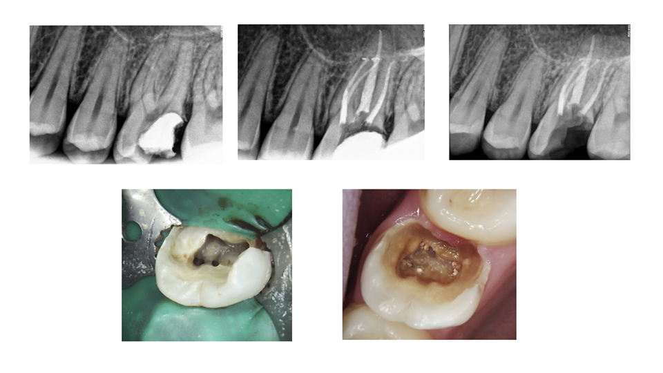
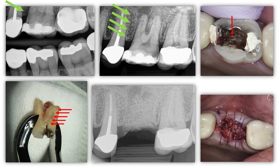
PERSISTENT PAIN FOLLOWING RCT: Determining the etiology of persistent pain following a “well-done root canal” can be a challenge as there are many possible explanations. Such possibilities include an untreated canal, coronal leakage, fracture, or referred pain from another tooth or non-odontogenic structure. The attached images are of a 41 year-old patient who returned to our office 5 months following RCT tooth #8. The patient described her “persistent pain” as tenderness when she palpates the facial gingiva overlying apex #8. CBCT examination demonstrated that tooth #8 is facially inclined with its apex located outside of the bony housing of the maxilla—allowing […]
Category: Diagnosis
TRAUMA AND CBCT
TRAUMA AND CBCT: Yet another trauma case where CBCT evaluation was an integral part of diagnosis and treatment planning. This patient presented to our office two days after taking an elbow to the mouth during a volleyball match. The patient stated immediately following the incident, he noted bleeding of the gingival sulcus and pain tooth #8. He denied tooth movement or malpositioning. Tooth #8 had a full coverage crown, was tender to percussion and palpation, Class III mobility, no response to cold, no isolated deep probing depths, and widened PDL space in the apical third. Teeth #6, 7, 9, 10, and […]
FAILING CHIN AUGMENTATION OR INFECTED RETENTION SCREW?
FAILING CHIN AUGMENTATION OR INFECTED RETENTION SCREW? Here is an interesting case with the following presentation… – 42 yo healthy female – Chief Complaint: Recent onset pain on the LR chin area – Hx of facial reconstruction including mandibular surgery to address significant Class II malocclusion – Chin augmentation also performed at that time; outlined by red arrows – CBCT shows the location of the chin graft with a radiolucency at the interface of graft and communication with root apices of #26 and #27 – Surgery done in 1989 – Recent onset discomfort associated with #26, 27 area with radiographic […]
Read More… from FAILING CHIN AUGMENTATION OR INFECTED RETENTION SCREW?
IMPORTANCE OF VITALITY TESTING
IMPORTANCE OF VITALITY TESTING: This 36-yo healthy, Caucasian female was referred for endodontic treatment of asymptomatic tooth #23. Radiolucency tooth #23 was noted by her dentist upon evaluation for Invisalign®. See chart for clinical findings. Pt denied history of trauma. Due to positive response to vitality testing, expansion of L cortical plate, tooth mobility, and mixed radiopaque-radiolucent radiographic appearance, the lesion was determined to be of non-endodontic origin. The patient was referred to an oral surgeon for further evaluation and treatment. We recently followed-up with the patient’s oral surgeon. The lesion was biopsied and determined to be fibro-osseous dysplasia. […]
PROPER DIAGNOSIS OF NON-RESTORABLE TOOTH FRACTURES
PROPER DIAGNOSIS OF NON-RESTORABLE TOOTH FRACTURES: This patient presents to our office with attempted excavation of an existing restoration and to evaluate the extent of a fracture in the crown (the patient had an MOD composite restoration). It was known by the restorative dentist that the tooth was necrotic (Note MB root lesion). For whatever reason, the dentist decided at this point to temporize and send to us for case management. BEFORE clinical exam and considering CBCT, we had noted several key factors in the PA and BW radiographs; the most important facto was the presence of bone loss on […]
Read More… from PROPER DIAGNOSIS OF NON-RESTORABLE TOOTH FRACTURES