ENDODONTICS AND IMPLANT DENTISTRY: […]
ENDODONTICS AND IMPLANT DENTISTRY
ENDODONTICS AND IMPLANT DENTISTRY: […]
C-SHAPE RETREATMENT – Conical second molars can have some of the most variable anatomy in the adult dentition. Here we present two “c-shape” molar retreatment cases that highlight this unusual morphology. Due to the twisting, merging, splitting, and curving of these canals which are often all be joined by a thin slit of tissue called an isthmus, meticulous endodontics is essential in order to achieve healing. […]

DENTAL ANATOMY: Here is an interesting one that came in today. Usually when we come across a 5 canal lower molar, it is usually in a first molar. Also, it will more commonly occur in first molars. Here we present a “5 canal” lower molar in a second molar, with the extra canal in the distal aspect as opposed to mesial aspect. Maybe not as cool since 2 of the 3 distal canals share one common exit each, but pretty photos nonetheless…all in the efforts to eviscerate pulp tissue… […]

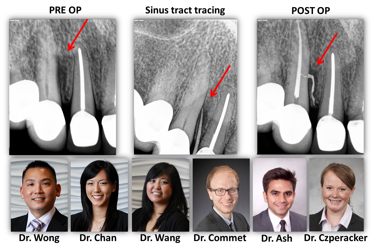
This just in!! Not your typical radix enomolaris root presentation….cool! […]

Both of these teeth were treated at our office at 2 different times. Tooth #3 (on the right) was treated today while tooth #2 (on the left) was treated previously. Taken together, there are 9 canals between the 2 of them!! Pretty cool cases…we would have like to negotiate one of the canals on tooth #3 (the MB2) as it was non-patent; however we still feel that this represents a very favorable endodontic result! […]

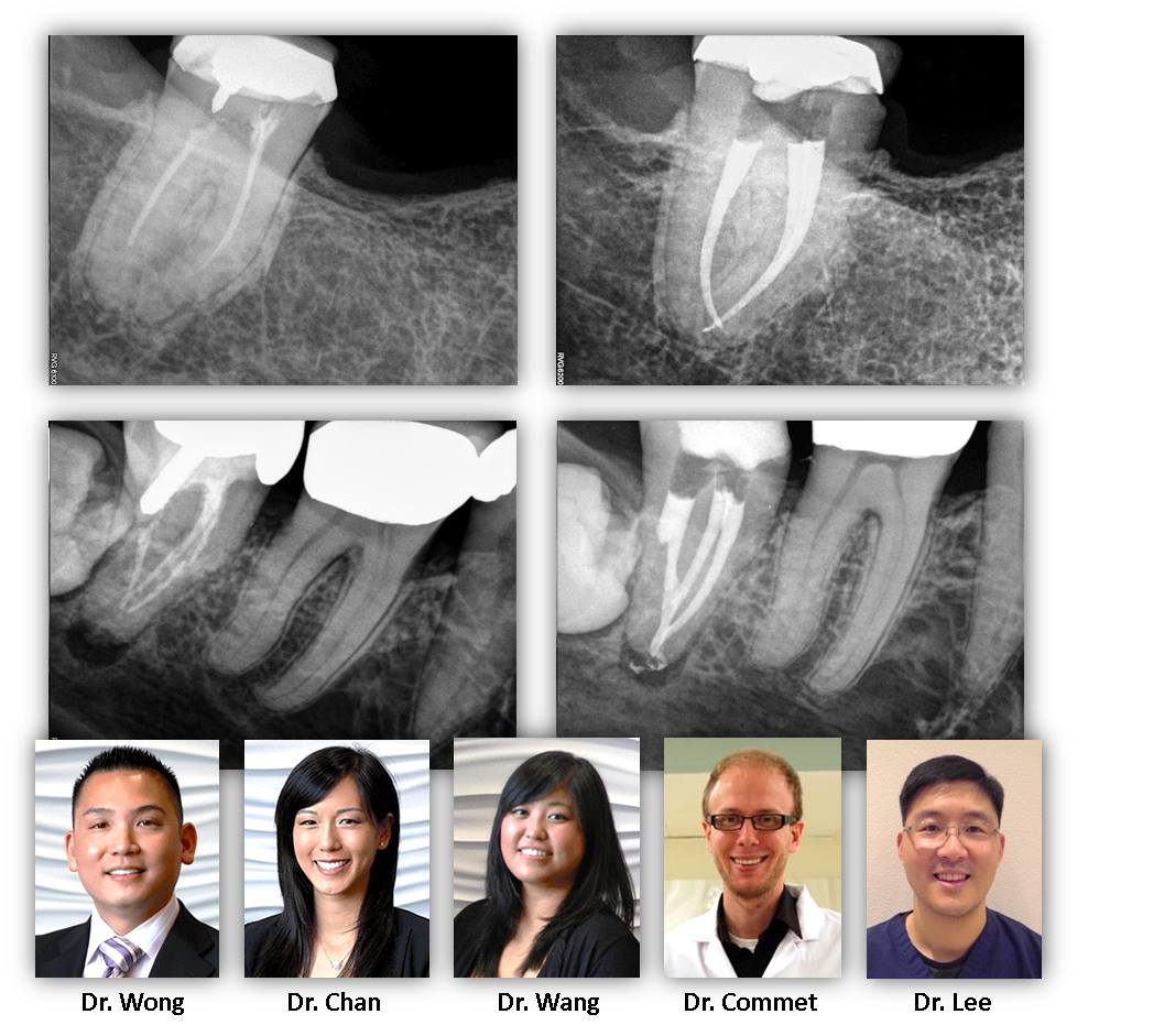
These teeth from two patients this week were initially started by the restorative dentist. After careful debridement, the dentists noted that there were likely additional canals and referred the patients to Renovo. Interestingly, these teeth actually had three canals that split and merge at various levels of the teeth. A favorable outcome is anticipated and these cases demonstrate the benefit and need for microscope-aided endodontics. Aside: Most endodontists actually find the mandibular premolar (rather than a molar) to be the most challenging tooth to treat due to the prevalence of this type of complex anatomy in a relatively confined space. […]
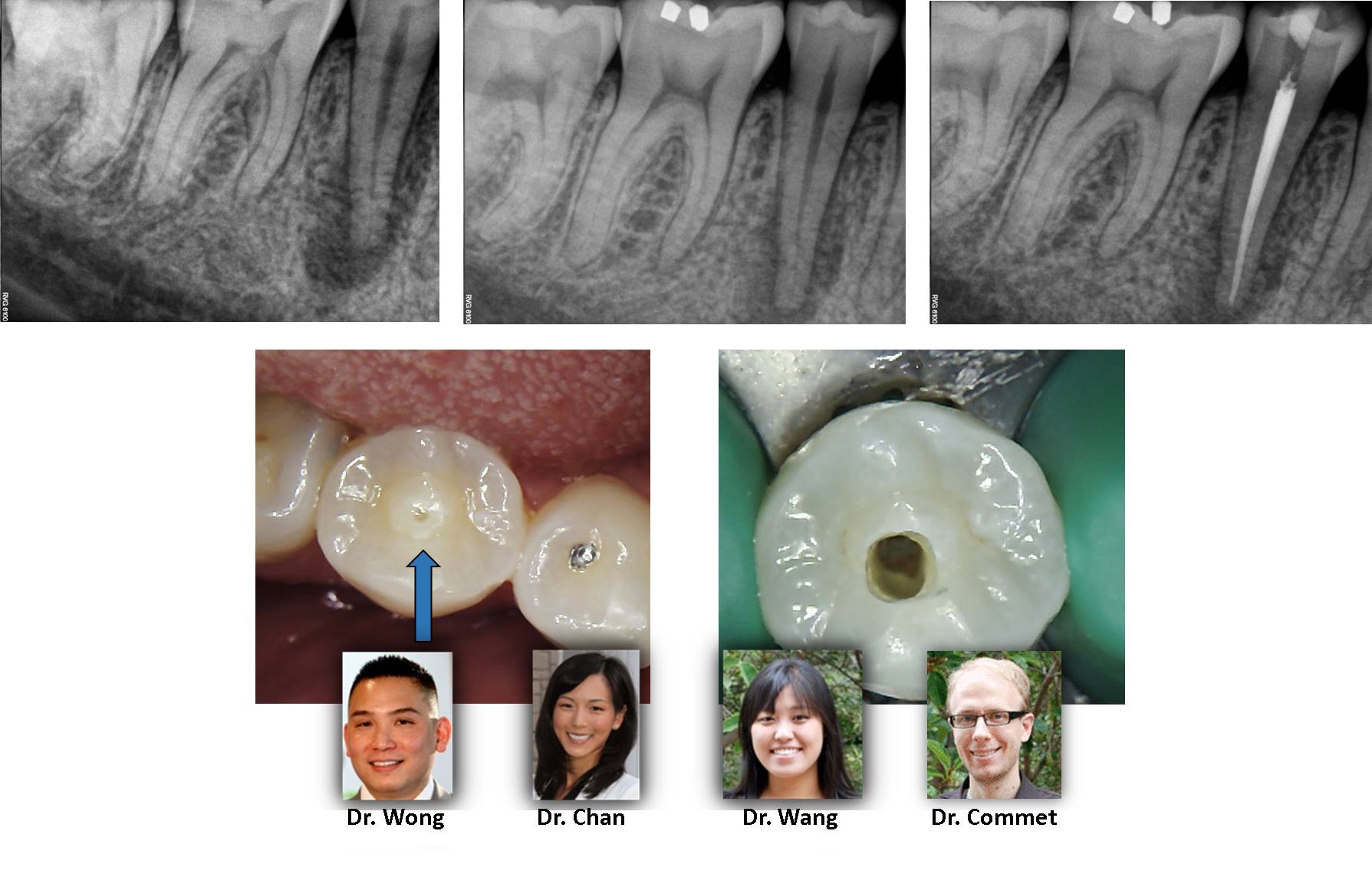
This tooth demonstrates the pitfalls of a dens evaginatus. As can be noted on the clinical photograph, the atypical central cusp quickly wears due to attrition resulting in pulpal exposure and corresponding necrosis. Careful inspection of the occlusal anatomy is important in these cases as the radiographic appearance is similar to that of a vertical root fracture but this tooth has a very favorable prognosis. […]

This case represents a rare anatomic variant of a tooth anatomy much more commonly seen in the mandible: “c-shape”. Because the maxillary variant often has fused roots with DB/P canals that join very early in the pulp chamber with a separate MB canal that joins near the apex, some refer to this rare tooth as having a “semi-colon” anatomy. Take a look at the clinical photograph; can you see the semi-colon? […]

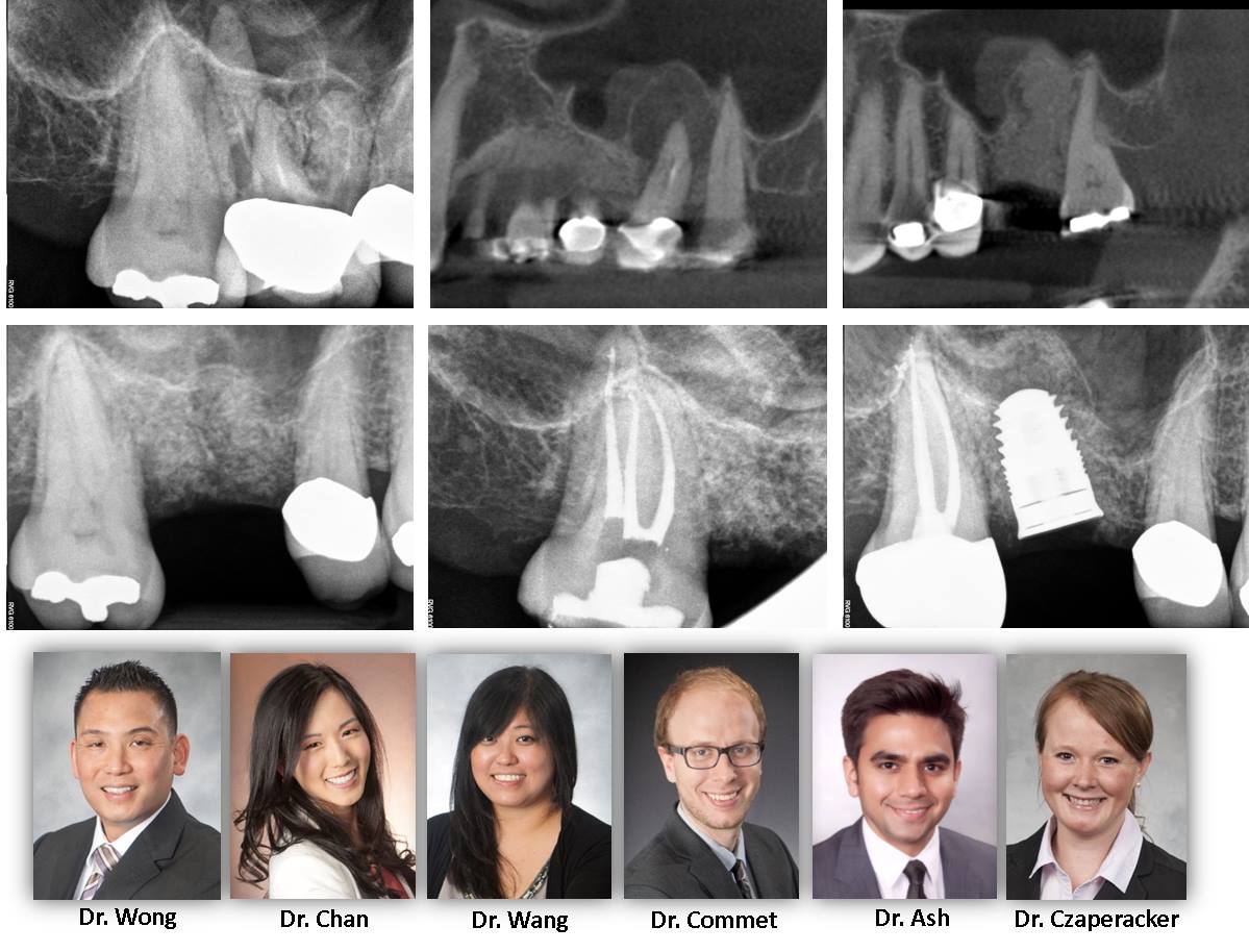
It is only Tuesday and here is a sampling of cases that have come through this week. We demonstrate bifurcations, middle mesial canals, radix entomolaris, significant curvatures. All of these are examples of anatomical considerations when rendering root canal therapy in order to maximize disinfection and long term success. Turkey Day is just around the corner!! […]