HOPELESS PROGNOSIS?: Based on the pre-operative PA, many dentists would suggest to their patients that this tooth has a poor or hopeless prognosis. Clinically it had a 12 mm probing to the apex and radiographically it has a large apical lesion that was confirmed to extend around the entire MB root on CBCT. Even more concerning was that the lesion was not well centered around the root apex and was localized around the lateral root surface. Frequently patients like this are told “This tooth is fractured, it has to come out” without even attempting endodontic treatment. This case presents a great […]
Category: Cool Cases
HOPELESS PROGNOSIS?
Emergency Tooth Replantation: Procedures, Success Rates & Risks
Emergency Tooth Replantation: Procedures, Success Rates & Risks
OROANTRAL COMMUNICATION CAUSED BY ODONTOGENIC PATHOLOGY
OROANTRAL COMMUNICATION CAUSED BY ODONTOGENIC PATHOLOGY
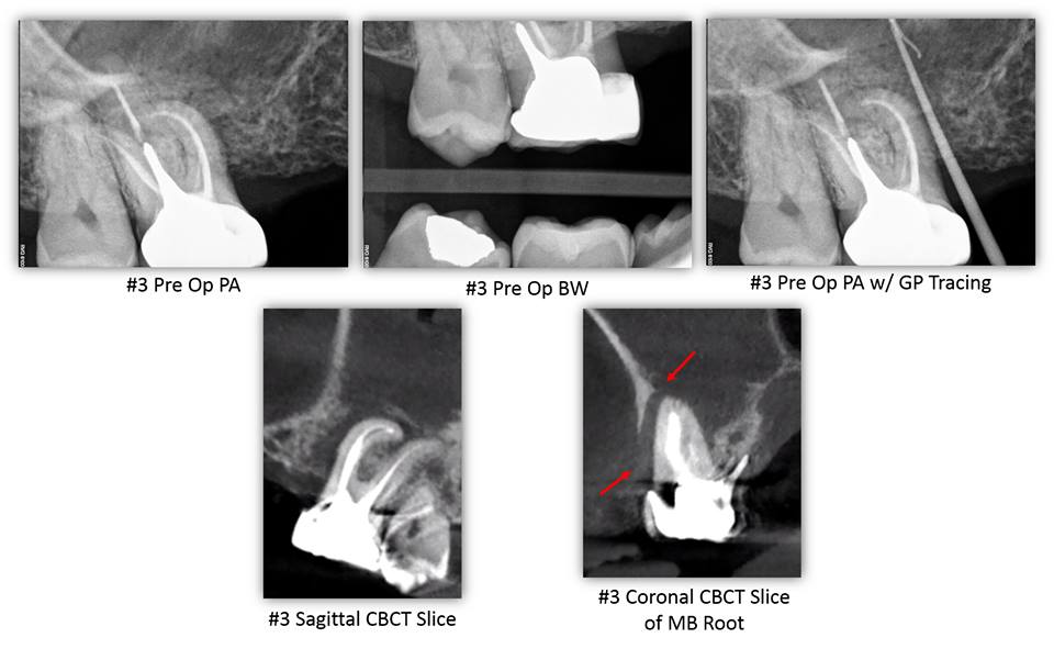
OROANTRAL COMMUNICATION CAUSED BY ODONTOGENIC PATHOLOGY– This patient presented for evaluation of tooth #3. His chief complaint was a painful swelling of gum that would sometimes bleed in the area of tooth #3. The patient reported RCT #3 was completed over 20 years ago by an Endodontist. The patient stated that he is congenitally missing teeth #4, 5, 12, and 13. The patient denies wearing an RPD. The patient’s chief complaint was consistent with a sinus tract located mesial to tooth #3 at the level of the alveolar crest. The sinus tract was traced with gutta-percha. Tooth #3 had an intact […]
Read More… from OROANTRAL COMMUNICATION CAUSED BY ODONTOGENIC PATHOLOGY
LAST CASE OF THE WEEK
LAST CASE OF THE WEEK

LAST CASE OF THE WEEK: Non-restorable #31 – Bridge sectioned between #29 and pontic #30 – #31 and residual root tip #30 extracted – Extraction site #31 grafted – Site #30 ridge augmented in preparation for dental implants to replace the existing FPD. […]
Another One Bites the Dust
ANOTHER ONE BITES THE DUST
ANOTHER ONE BITES THE DUST: I was actually thinking for moment to try and save this one…but the cost:benefit analysis for retreatment, perforation repair, new indirect restoration just didn’t seem worth it. Additionally with that much retention needed in the form of the post/core), there is surely a significant compromise in the lack of good tooth structure left to work with. We decided to go with the more predictable route here. […]
Thermafill retreatment #30 with some accessory portals
Thermafill retreatment #30 with some accessory portals

Thermafill retreatment #30 with some accessory portals. Root canal completion #31. More anatomy….. […]
Read More… from Thermafill retreatment #30 with some accessory portals
Lesion Shape Suggestive of Root Canal Anatomy
LESION SHAPE SUGGESTIVE OF ROOT CANAL ANATOMY
LESION SHAPE SUGGESTIVE OF ROOT CANAL ANATOMY: This is a pretty good demonstration of periapical lesion associated with the canine tooth (#6). Pre-operative radiographic survey reveals that the lesion is off to the side at the apical third. Post-operative radiograph highlights a couple accessory canals providing a route of infection and inflammatory response in the area of the lesion. Good demonstration of anatomy here. […]
Read More… from LESION SHAPE SUGGESTIVE OF ROOT CANAL ANATOMY
Double Curves
DOUBLE CURVES

DOUBLE CURVES: These are a couple recent cases demonstrating double curves at the apical third of the roots. Sticking to sound instrumentation techniques are really helpful in cases like these to avoid separating files within the canal system. […]
An Unusual 'Post' Removal
AN UNUSUAL ‘POST’ REMOVAL
AN UNUSUAL ‘POST’ REMOVAL Apicoectomy can be a highly predictable treatment option for patients with failing endodontic therapy. Modern microscurgery involves not only root resection, but also retropreparation using ultrasonics and retrofil using bioceramic materials like MTA. Without these two key factors, treatment success drops from ~87% to nearly 60% and is the reason apicoectomy tends to have a “50/50” connotation with some oral surgeon offices. The following is a great case to demonstrate the possibilities in modern endo! Pre-operatively you can note that the ‘post’ (silver point) goes to the apex and CBCT showed a corresponding lesion. Placing bioceramics on […]
Broken Instruments...or Modified Root Filling?
BROKEN INSTRUMENTS…OR MODIFIED ROOT FILLING?
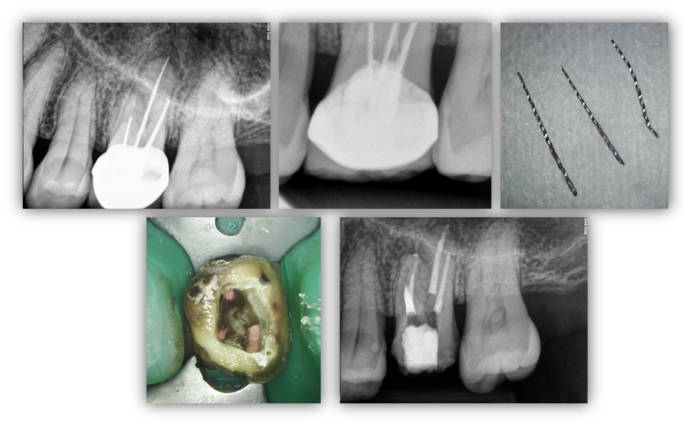
BROKEN INSTRUMENTS…OR MODIFIED ROOT FILLING? Here we present a relatively interesting case, #14, with a recurrent infection associated with the MB root. Other than pain associated with this tooth, medical history remains unremarkable. We were able to remove the “root filling” which appears to be endodontic instruments that may have also been used in initial instrumentation. In any case, we were able to remove the all three instruments and locate a previously untreated MB2 canal. This would prove to be significant as this it the only canal in the MB root that was able to be instrumented to full root […]
Read More… from BROKEN INSTRUMENTS…OR MODIFIED ROOT FILLING?